


3D Pollu Skin
A FAIRE The first worldwide 3D Full-tickness skin model
including sebaceous glands
What about?
Current skin tissue engineering technologies often only include the epidermal components and remain partially effective in their ability to restore the complexity of the dermal compartment, particularly skin appendix.
Nowadays, studies about sebaceous glands rely mainly on the use of 2D cell culture based on immortalized cell lines and most 3D models are ex-vivo isolated sebaceous glands with limited shelf life.
To overcome these limitations, LabSkin Creations designed an unique model by using its porous scaffolding biomaterials to support cell growth and 3D organisation of sebaceous glands in 3D.
A 3D Full-thickness skin model containing hIPS-derived Sebocytes
3D SEBOSkin model integrating hIPSC-derived sebocytes are morphologically consistent with a well-organised and terminally differentiated epidermis and presents a cohesive dermal-epidermal structure.
After 48 days of culture, dermal fibroblasts seeded with hIPSC-derived sebocytes were able to colonize the whole collagen-GAG-chitosan porous scaffold and neo-synthesize their own extracellular matrix.
hiPSC-derived sebocytes seeded within LabSkin porous scaffold self-organize into well-defined 3D organoids embedded into extracellular matrix neo-synthetized by human fibroblasts.
More importantly, 3D SEBOSkin actively accumulated lipid droplets demonstrating their functionality.
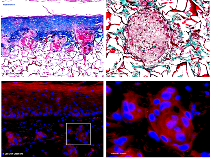
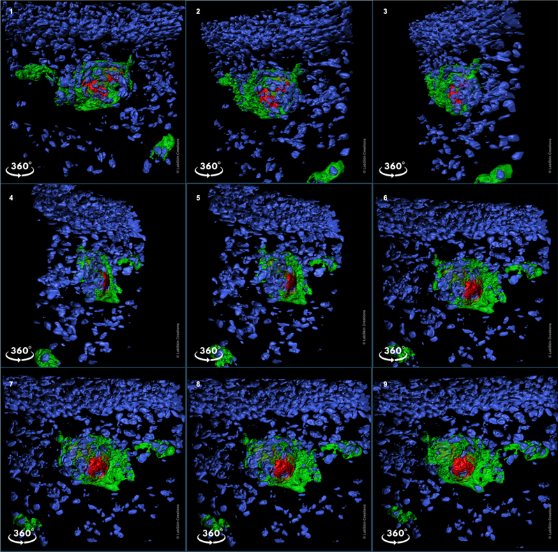
“3D within 3D” Well-defined 3D organoids embedded into a 3D skin equivalent
Sebaceous glands are grossly identifiable by their “foamy” appearance on microscopy. SEBOSkin shows a typical glandular structural organization as observed in vivo with relatively undifferentiated sebocytes located along with the outer layers of the gland, gradually becoming more differentiated and filled with lipid products toward the center.
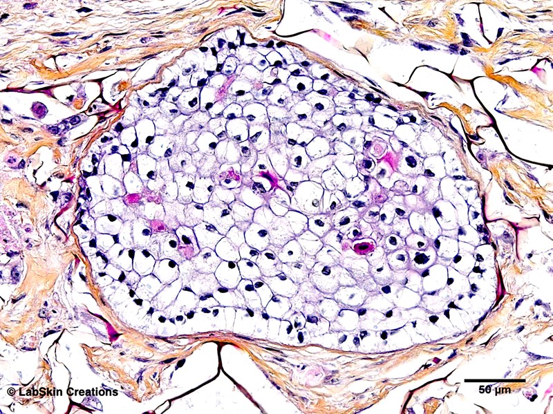
Here, fully mature sebocytes have pyknotic nuclei, indicative that they are about to degenerate to form the necrosis zone.
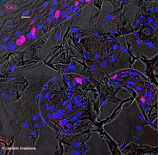
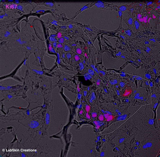
As observed in vivo, Ki67 staining was restricted to the basal cells surrounding the 3D organoids, and was not apparent in the differentiating hiPSC-derived sebocytes.
SEBOSkin expresses key sebaceous gland markers
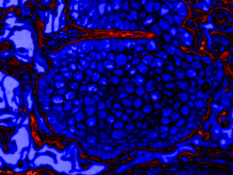
Similarly to normal human skin, 3D SEBOSkin model expresses both specific sebaceous differentiation markers MUC1 and CK7.
The spatial distribution of MUC1 is confined to the centre of the gland and surrounded by CK7, demonstrating the centripetal maturation of the hiPSC-derived sebocyte organoids in the scaffolding biomaterials.
3D SEBOSkin - A fully functional model
To assess the functionality of our 3D model, the skin equivalent samples were treated with different concentrations of arachidonic acid, well known to stimulate the production of intracellular lipids of sebocytes.

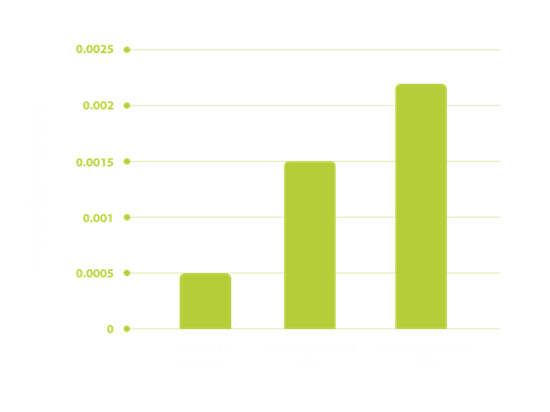
Arachidonic acid increased significantly squalene expression in our 3D skin model containing iPSC-derived sebocyte in a dose dependent manner, compared with the untreated control condition demonstrating its functionality.
By combining LabSkin advanced scaffolding biomaterials made of collagens, GAG and chitosan optimised in composition, proportion and porosity with innovative cell culture technologies, LabSkin Creations developed the first worldwide 3D full-thickness skin model including sebocyte organoids derived from human iPSC.